Arterial cannulation procedure follows the technique of insertion of cannula inside the artery for the sampling and blood pressure monitoring
Arterial line: Indications
Continuous blood pressure monitoring in shock and critically ill patients especially in the presence of marked peripheral vasoconstriction.
Frequent arterial blood sampling.
ABG- Arterial blood gas analysis
The technique of radial artery cannulation
The radial artery is the commonest site for arterial cannulation
Gently dorsiflex and secure the wrist across a pad, or folded towel.
Clean the area with povidone-iodine or any antiseptic solution.
Arterial cannulation procedure
Palpate the radial artery over the wrist. 1% lignocaine solution injected intradermally using a 26 g needle. This anesthetizes the area and prevents arterial spasms at the time of puncture.
Insert a 20-gauge cannula at a fairly steep 30-degree angle to the skin. When the stylet pierces the vessel and blood begins to flow into the stylet hub, decrease the angle of the catheter to about 10 degrees.
At this point, carefully advance the stylet another ½ millimeter to ensure that the catheter tip is in the lumen of the vessel.
Then gently withdraw the stylet and advance the catheter into the artery. Under no circumstance, the catheter advanced against resistance in the artery.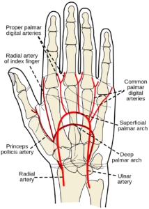
In the presence of hypotension, the radial pulse is often not palpable. In such circumstances, the femoral artery can be cannulated using a long 20-gauge cannula.
Invasive arterial pressure monitoring
The cannula connected to a pressure tubing, which is attached to a transducer. The transducer connected to a monitor for continuous display of waveforms and blood pressure.
Modified Allen’s Test.
The test does check the presence of collateral blood supply and identify patients at high risk for ischemic complications during or after radial artery cannulation.
The patient closes his hand as tightly as possible for a period of one minute in order to squeeze blood out of the hand. The examiner compresses the wrist between his thumb and fingers, thus occluding the radial artery. The examiner then releases pressure over the ulnar artery while still maintaining pressure over the radial artery and the patient asked to extend his fingers.
The return of color to the hand and fingers is noted. Normally, full blushing is complete at 7 seconds, 8 to 14 seconds is borderline, and 15 seconds is considered abnormal. If the ulnar flow is sluggish, then the poor ulnar collateral flows better to avoid radial cannulation.
For Arterial cannulation custom made arterial cannula available with polyurethane material specialized for this procedure