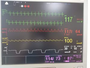
The monitor provides continuous, non-invasive, automatically calibrated measurements of both functional oxygen saturation of hemoglobin and pulse rate.
The instrument combines the principles of spectrophotometric oximetry and plethysmography. It consists of an electro-optical sensor that applied to the patient and a microprocessor-based monitor that processes and displays the measurements.
The electro-optical sensor contains two low-voltage, low-intensity light-emitting diodes (LEDs) that serve as light sources and one photodiode as a light receiver. One LED emits red light (approximately 920 nm.)
How to measure spo2 level: Principle behind that
When the light from the LEDs transmitted through the blood(arterial) and the various tissue components, a portion of it absorbed. The photodiode in the sensor measures the light transmitted, and this measurement used to determine how much light absorbed.
With each heartbeat, a pulse of oxygenated arterial blood flows to the sensor site.
This oxygenated hemoglobin differs from deoxygenated hemoglobin in the amount of red and infrared light that it absorbs.
The control panel measures the absorption of both red and infrared light and uses those measurements to determine the percentage of functional hemoglobin that is saturated with oxygen.
Discussion
Initially, light absorption determined, when the pulsatile blood not present in the blood pool. This measurement indicates the amount of light absorbed by tissue and non-pulsatile blood absorption that does not change substantially during the pulse.
This is analogous to the reference measurement of a spectrophotometer.
Absorption is then measured when the pulsatile blood is present.
In that measurement, light absorption at both wavelengths, changed by the presence of the pulsatile, arterial blood. The corrects the measurement obtained during the pulsatile flow for the amount of light that was absorbed in the initial measurement.
The ratio of the correct absorption at each wavelength
then used to determine functional oxygen saturation. Each sensor is checked at the time of manufacture.
FUNCTIONAL v/s FRACTIONAL SATURATION
As this machine measures functional oxygen saturation, it may produce measurements that differ from the instruments measuring fractional oxygen saturation.
Functional oxygen saturation is characterized as oxygenated hemoglobin communicated as a level of the hemoglobin that is equipped for transporting oxygen.
The method uses two wavelengths to measure saturation, it measures only oxygenated and deoxygenated (i.e. functional) hemoglobin.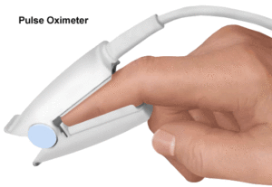
In contrast, some other laboratory instruments report fractional oxygen saturation values. Fractional saturation defined in which oxygenated hemoglobin expressed as a percentage of all hemoglobin, whether or not that availability of hemoglobin for oxygen transport.
Measured dysfunctional hemoglobins included in this calculation. Consequently, when measurements from the oximeter compared with those from another instrument. It’s important to determine whether other instruments measuring functional or fractional saturation.
Those measurements converted to functional, using the following equation :
Functional saturation= fractional saturation * 100/100%- [%carboxyhaemoglobin + % methemoglobin]
MEASURED CALCULATED SATURATION
When oxygen saturation calculated from a blood gas measurement of the partial pressure of arterial oxygen (PaO2). The calculated value may differ from the oxygen saturation measurement of the oximeter.
Oxygen saturation value calculated from blood gas PaO2 not necessarily correctly adjusted for the effect of variables that shift the relationship between PaO2 and oxygen saturation.
These variables include temperature, pH, the partial pressure of carbon dioxide (PC02), and the concentration of fetal hemoglobin.
At the point when oxygen saturation determined from a blood gas estimation of the partial pressure of arterial oxygen (PaO2), the determined worth may vary from the oxygen immersion estimation of the oximeter.
This is on the grounds that an oxygen immersion esteem that has been determined from blood gas.
PaO2 not accurately balanced for the impact of factors that move the connection between PaO2 and oxygen saturation.
SPO2 SENSORS
Warning
Use only L&T approved sensors. The use of sensors produced by other manufacturers
may result in improper oximeter performance. Incorrect application or use of an improper sensor may cause tissue damage or improper operation. Carefully read the “Warning” section of this manual and the directions for use provided with the sensor.
SELECTING A SENSOR
Each sensor designed for application to a specific site(s) on patients within a designated weight range.
To select the appropriate sensor, consider the patient’s weight and which sensor application sites available
Level of patient activity, whether sterility required, the anticipated duration of monitoring, and the adequacy of the patient’s perfusion.
Adult probe for adults above 40 Kg.
Infant probe for patients above 1 Kg. to 80 Kg.
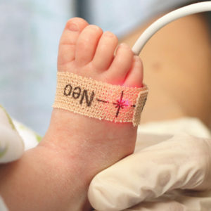
Newborn probe for patients above 40 Kg. and below 3 Kg.
Prepare the application site, remove nail polish, the clean surface area of contact in case of neonates, and apply the oxygen transducer using the sensor application guide of pulse oximetry transducer.
The perfusion indicator fills up the bar and plethysmographic waveform display accompany by an audible beep and numerical values of SpO2 and PR (pulse rate) displays on the screen.
PR displays in yellow color to illustrate the source from pulse oximetry.
The spo2 sensor comprises of two parts :
(a) Patient end (Main Sensor)
(b) Monitor end (Extension Cable)
Do not connect the SpO2 sensor and non-invasive blood pressure cuffs to the same limb of the patient.
Performance consideration
To ensure optimal performance, use an appropriate sensor. Apply the sensor as per the manufacturer’s instructions. Keep the sensor site at the level of the patient’s heart and
observe the Warnings and Cautions indicated in the sensor’s booklet
Instructions booklet
If the excessive ambient light is present, cover the sensor site with opaque material. Light sources that can affect performance include surgical lights, especially those with a xenon light source, fluorescent lights, infrared heating lamps, and direct sunlight.
In case of the disturbed waveform (if the waveform is noisy or the patient is unstable), evaluate the following things: Check whether the sensor is properly placed on the patient.
Use a new sensor with fresh adhesive backing.
Move the sensor to a more stable site.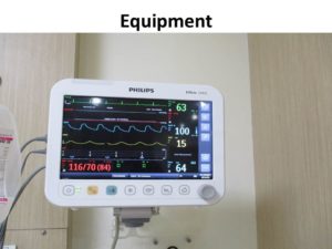
Though the console designed to minimize any effects of electrosurgical interference, such interference gives a problem, perform the following actions:
Move the cables of 6 am and the Cautery unit as far as possible.
Plug the Cautery unit into the different mains socket. Move the sensor away from the Cautery site and ground plate as much as possible.
Check whether the sensor is dry and firmly attached. Avoid crisscrossing of Cautery cables with monitor cables
Accessories for Pulse Oximeter from the probe for adults application