Percutaneous Tracheostomy a surgical opening of trachea while tracheostomy creation of stoma at the skin surface, which leads to the trachea. It may be used as temporary or permanent, according to the conditions of the patients. The procedure did with Dilating forceps, guidewire in tracheostomy kit
Indication.
Upper airway obstruction
Swelling resulting from burns
Prolonged ventilation
To provide broncho pulmonary toileting and to protect the airway
Conversion of cricothyroidotomy
An alternate pathway of breathing bypass
The upper airway obstruction
Improve alveolar ventilation
Tracheostomy -Congenital
Laryngomalacia(congenital disease)
Vocal cord paralysis
Inflammatory
Acute epiglottis
Acute Laryngo-bronchitis
Diphtheria
Traumatic injury
Malignant conditions
Bilateral abductor paralysis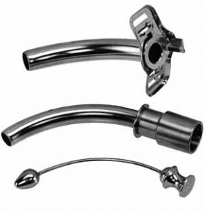
Foreign bodies
Bronchial asthma
Strider
Types of according to the uses
1.Elective
2.Permanent
3.Emergency
Effect of a tracheostomy.
The larynx is bypassed.
There is a risk of respiratory tract infection.
Foreign body reaction can occur causing local inflammation.
Principle
Consent
Before getting consent from the patients and patients guardians, you must have explained about the precaution, procedure, risk, and complication clearly.
Before the procedure confirms with the ultrasound, whether there is a presence or absence of large blood vessels
Nasogastric feeding should be stopped at least two hours, that may reduce any chance of aspiration of gastric content to the lungs.
Sedation, analgesia drugs, muscle relaxant, and vasoconstriction drugs kept ready before the procedures.
oxygen supply with the appropriate device for delivery with a connector. keep the suction ready.
Bronchoscope for visualization of the bronchus and carina if needed.
It is done under local anesthesia
The percutaneous tracheostomy technique is new and minimally invasive procedures.
The rapid method of insertion with less complication as compared to other conventional methods
The Seldinger’s techniques to guide the specially designed guidewire dilating forceps into the trachea, which then dilates the trachea
Positions
Patient in the supine position with neck extended and a wedge keeps a roll under the shoulder.
Tracheostomy Procedure
Before incision of the injection of the 1% lidocaine with 1:20000 epinephrine.
lubricate the tracheostomy tube 2% lignocaine jelly.
The use of an inner cannula reduces the airway size by 1.0 mm
A transverse skin incision overlying the upper trachea, that separating the strap muscle of the neck in the midline by retractors
Often thyroid isthmus may displace or divided to allow for adequate exposure of an anterior surface of the trachea.
The tracheostomy tube placed through the second or the third tracheal ring.
keep the suction ready and oxygen by the side.
After incision inserts the tracheotomy tube with the guidewire or an obturator is take out and suction is done when there is a secretion.
Inflate the cuff and check for the position of the tracheostomy tube with an assessment of chest movement, auscultation after confirmation sees the capnography for the confirmation chest, x-ray must be taken when there is extended pneumothorax.
Fix the tube with Silk suture at both sides of the flange and attach the tag tie to wind around the neck for support.
Tracheostomy Instrument kit
Kit contains mainly scalpels 14 G Cannula
10 CC syringe
Guidewire with introducer
A dilator with different sizes
Guidewire dilating forceps
Suction control valve
Tracheostomy tube and obturator with the lumen
Complication
Tracheal stenosis
Tracheoesophageal fistulas
Patient discomfort
Tracheal trauma (bleeding)
Tracheo-innominate artery damage