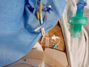
The procedure mainly used for the continuous central venous pressure CVP monitoring and for purpose of the fluid management, drug therapy, etc. central venous catheter is placed either in the internal jugular vein or subclavian vein, femoral vein, axillary vein.
CVP Indication
Infusion and fluid volume therapy
Patients requiring CVC for
Hemodynamic monitoring
Administration of special medications – vasopressors, antibiotic drugs, correction of
electrolytes, chemotherapy, etc
Access in patients where peripheral access is not possible
Total parenteral nutrition
Central venous pressure normal 7 to 12 mm Hg
For the administration of the high osmolarity solution
Intermittent central venous pressure monitoring
for venous blood sampling
for the no peripheral venous access
patients with injured extremities
for the shock patients
The material used in CVP
Seldinger needle or valve needle
Guidewire with length marking flexible J- tip /straight tip made of nitinol
Scalpel 11 sizes with BP handle
Dilator
Double lumen /triple lumen made of an opaque catheter made of polyurethane with a soft tip
With valves and three-way adapters
Colour coded Luer lock connection
Fixation wings for fixing the catheter
Luer lock syringe
Ultrasound sterile jelly
Drapes for the site (center hole )/normal drape
Tegaderm /condom or sterile gloves for the ultrasound probes
Drape for the ultrasound cable
Sterile solution for the skin preparation and alcohol-based disinfectant.
The appropriate size of suture for the catheter fixation
Tegaderm dressing or custom dressing for the CVP
Colour coding used in the CVP
Yellow color – CVP monitoring
Red color
White color
Blue color
5 ml syringe
secondary fixation wings
Procedure
The site chosen for insertion would depend on the clinical situation. Although subclavian lines are superior in terms of incidence of catheter-related bloodstream infections, in an unstable patient, it is preferable to use the internal jugular vein because of the higher probability of serious complications with the subclavian route.
If there is a likelihood of severe coagulopathy or very low platelet count, (before these parameters are known) the femoral site may be used.
Central line insertion is very rarely done as an emergency procedure – if you need to use inotrope/vasopressor for support of blood pressure, it is OK to use a peripheral IV line.
so, until you are able to get central access. Remember, stabilization of the patient is a priority -line insertion is not. Hence, a CVC should not be inserted without taking appropriate aseptic precautions. Do seek help from senior colleagues if you are not sufficiently experienced with the procedure
Contraindications
Inflammation of the skin at the site of puncture anatomical anomaly
Very severe pulmonary emphysema
Postoperative changes in the site area
Some of the Catheter coated with Rifampicin and Miconazole contraindicated to known hypersensitivity to following drugs.
Risks in Central Venous Pressure Procedure
CVP monitoring finds the correct position of the line in situ
Hematoma at the site of puncture
Catheter sepsis
Pneumothorax or chylothorax
Haemothorax
Infusion hydrothorax
Incorrect catheter positions
Cardiac arrhythmias due to the incorrect intracardiac placement of the catheter
Arterial rupture
Endocarditis due to mechanical irritation
Arterial injuries due to incorrect puncture
Catheter-induced thrombosis
Thromboembolism
Thrombophlebitis of superior vena cava
Thoracic duct injuries
Damage to brachial plexus
Damage to the phrenic nerve
Warning
Do not reuse the single-use materials
Use strictly aseptic precaution & techniques for the CVP.
Position of the patients in Trendelenburg
Use Universal Precaution to all the patients against the Needlestick injury and contact with blood.
Complication
Vessel wall perforation
Cardiac tamponade
Hemorrhage
Pleural and mediastinal injuries
Air embolism
Catheter embolism
Nerve Lesion
Septicemia
Bacteraemia
Thrombosis
Arterial puncture
Infection
Removal of CVP Catheter
To remove the catheter needs the special precaution and attention to do.
Keep ready with the 11 size blade and sterile gauze pieces
Custom dressing material
First, remove the suture from the fixation site and Withdraw the catheter from the insertion site
After removal of the catheter apply pressure over the vessel to prevent hematoma
Apply custom pad dressing on the site of insertion with pressure
It recommended removing the catheter within 30 days of insertion.
After removal send it for culture for contamination
Discard it as per the recommendation of the Biomedical Waste management